User Interface
To enable users to interact with the device effectively, organisations develop a user interface (UI) that facilitates tasks such as uploading images, viewing reports, and utilising the device's features. Under IEC 62366-1, the integrator (the information-technology professional configuring and rendering the device's outputs into the integrating healthcare system) is an intended user of the device; this Installation Manual is the volume of the Instructions for Use directed at that intended user.
To help integrators in the process of developing a user interface, and to comply with the requirements regarding the information supplied with the device (Annex I, Chapter III of the MDR 2017/745), this section outlines the key elements of the user interface, their purposes, and how they assist healthcare professionals (HCPs) in making informed decisions about patient care. Clauses in this section that begin with "the integrator must" impose mandatory integration requirements; all other guidance in this section is recommended practice.
The user interface is divided into three main sections:
- Instructions for capturing images.
- Reports generated by the device.
- Label for regulatory compliance.
Each section plays a crucial role in enhancing the user experience and should be designed for clarity, ease of use and regulatory compliance.
Instructions
This section provides guidelines to help users capture high-quality images of skin lesions. Clear and simple instructions help users capture images and contribute to the quality of the experience and the data.
The requirements of this section apply to whoever captures the images, whatever that user is shown of the device's output afterwards. Together with the display of image quality to that user, described under Image Quality, they control the risk that an unsuitable image is analysed.
Select Problem Type
Instructions vary depending on the type of condition. For instance, localized lesions require different guidance compared to other conditions. Users can navigate to the relevant instructions by clicking a button or link.
Type of issue
Select the type of issue

Localized Lesion
For example, a mole or a pigmentation.
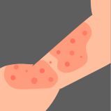
Something else
Any problem that is not on a fixed spot.
The integrator must let the user who captures select the type of problem before capture, so that the instructions shown are those for that type. This requirement controls the risk that an image is captured following the instructions for a different type of condition.
Instructions for Localized Lesions
Localized lesions, such as moles, require close-up images where the lesion is the focal point.
The integrator must show these steps to the user who captures images of localized lesions, at the moment of capture:

Capture the photo with your mobile phone.
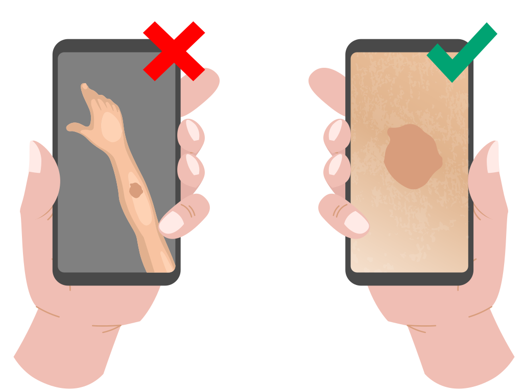
Center the photo on the lesion.

Take up to three images of the same lesion.
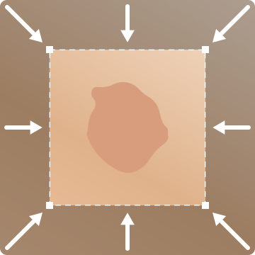
Crop the image.
For dermatoscopic images, specific instructions are unnecessary as the dermatoscope is designed to capture close-up, standardized images of skin lesions.
Instructions for Other Conditions
Conditions such as skin infections, inflammatory diseases, or rashes require different instructions due to their variability.
The integrator must show these steps to the user who captures images of other conditions, at the moment of capture:
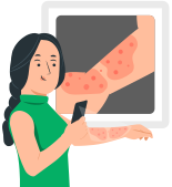
Capture the whole affected area with your mobile phone.
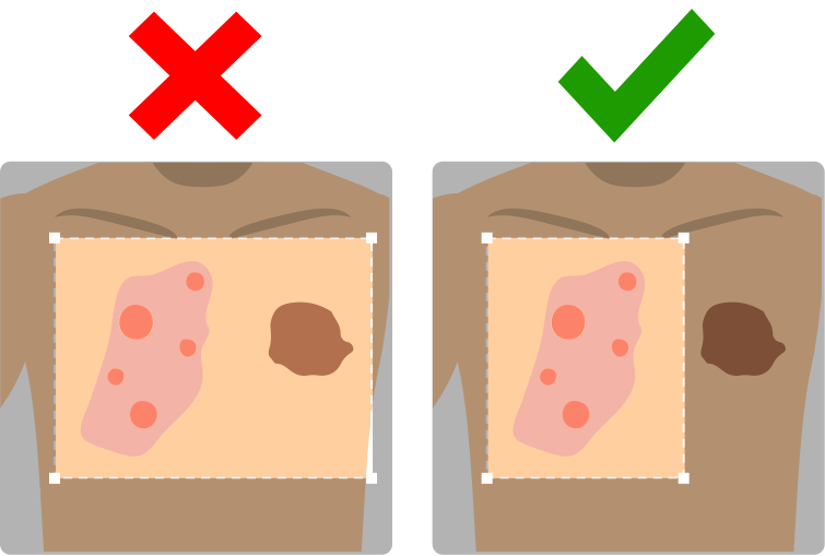
Only send one dermatological problem at a time.
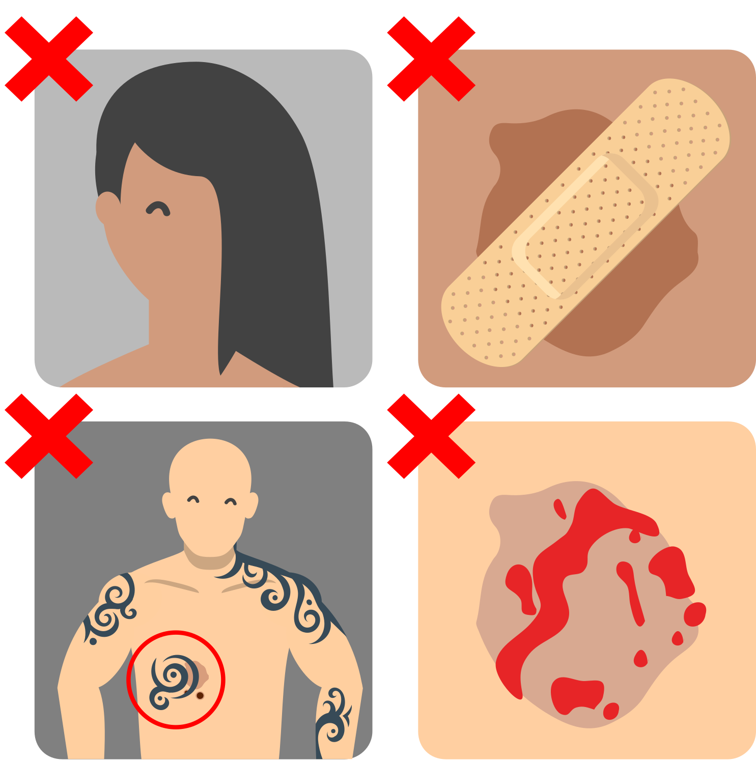
Avoid covering the affected area with clothing, hair or accessories.
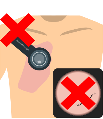
Do not use a dermatoscope. Take the photo with your mobile phone's camera.
Image Cropper
Cropping lets the user who captures frame the lesion, so that the affected skin fills most of the image and the background around it is left out of the analysis.

The integrator must provide an image cropper with which the user who captures selects the area of each image that contains the lesion or the affected skin, before the image is sent. The cropper must work in the four stages below, on every screen size. This requirement controls the risk that an image in which the lesion fills only a small part of the frame is analysed.
- Upload. When the user adds an image, the cropper shows it in full, with a control to remove it and, on a screen narrower than 1,200 pixels, a control to crop it. The control to remove the image stays available at every stage, and it discards the whole image, not the crop, so that the image can be captured again.
- Initial frame. The cropper places a crop frame on the image on its own, centred and covering 95% of its width, so that the user sees the edges of the crop before adjusting it. The frame cannot be cleared, so a selection always exists.
- Adjustment. The user drags the frame to move it and drags its corner handles to resize it, until the frame contains the lesion with a margin of the surrounding healthy skin. The frame has a free aspect ratio and a minimum size of 100 by 100 pixels on screen. The corner handles must be easy to grab with a mouse and with a finger: on a touch screen, the area that responds to a touch on each handle is at least 24 by 24 pixels, the minimum target size of success criterion 2.5.8 of the Web Content Accessibility Guidelines 2.2, even where the visible handle is smaller.
- Confirmation. The user confirms the crop. On a screen narrower than 1,200 pixels, the user saves the crop, and the cropper shows the cropped image, which the user can crop again or remove. On a wider screen, releasing the frame confirms the crop at once, and the user can adjust it again at any time before the image is sent.
The integrator must also:
- Apply the orientation recorded in the image before showing it in the cropper, so that the crop is taken in the orientation the user sees.
- Keep the original image until the image is sent, so that the user can crop it again from the original.
- Send the device the cropped image. The device receives an image, not crop coordinates.
- On screens narrower than 1,200 pixels, tablets included, show the image as a preview until the user chooses to crop it, open the frame only then, and close it when the user saves the crop.
Report
The sub-sections below define both presentation guidance and the UI elements whose implementation is mandatory. The mandatory report elements are the Top-5 prioritised differential view, the malignancy-prioritisation gauge, the referral recommendation, the image quality, the entropy, and the severity assessment wherever the device produces it. Each mandatory requirement exists to control a clinical risk arising from how the device's output is presented to the healthcare professional. The remaining elements below (the binary safety indicators, metadata, quick insights) are recommended integration features whose adoption is at the integrator's discretion.
The thresholds in the sub-sections below are suggested ranges. The right threshold depends on the use case, which is why this section characterises ranges rather than a single cut-off: the organisation sets the thresholds that suit its population and its clinical management protocol.
Integrators that do not meet these mandatory display requirements operate the device outside its intended use.
Full Diagnostic Report
Interactive
An interactive report combines various components into a visually engaging interface. This format is ideal for web or native applications, whether desktop or mobile.
Left arm
12/02/2026, 09:41:12
The normalized entropy value is high (69%), meaning the algorithm has low certainty about its analysis. Please keep this in mind when interpreting the results.
Select a condition
The algorithm has detected the following conditions.
- Condition A67.51%
- Condition B26.24%
- Condition C1.19%
- Condition D0.99%
- Condition E0.53%
Select other condition
Suspicion of malignancy
62%
Predicted by the algorithm
Report information
Information about the report.
- TimestampFeb 01, 2024, 10:06 PM
- Analysis performed in0.65 seconds
- Image modalityDermatoscopic
- Visual image quality82%
Severity Score
Automatic Psoriasis Area and Severity Index
Mild
Desquamation
Moderate (2)
Erythema
Moderate (2)
Induration
Mild (1)
Affected area
30% (2)
Severity
(PASI)
Time
Medical Device
Legit.Health Plus
Computational software-only medical device leveraging computer vision algorithms to process images of the epidermis, the dermis and its appendages, among other skin structures, providing an interpretative distribution of possible ICD categories and quantifiable data on the intensity, count, and extent of clinical signs.
AI Labs Group S.L.
BAT Tower, Gran Vía 1, 48001, Bilbao, Biscay (Spain)
UK Responsible Person
Apotech Consulting Limited
71-75 Shelton Street, London, WC2H 9JQ, England, United Kingdom
(01)8437025550005(10)1.1.0.0(11)20260123
1.1.0.0
2026-01-23
CE marking per Regulation (EU) 2017/745
Caution: consult accompanying documents
Read the electronic instructions for use (eIFU)
ifu-eu-mdr.legit.health
Interactive reports are highly recommended as they help users understand the output in a clear and compelling manner.
Plain Text
A plain text report integrates UI components into a straightforward format, suitable for older systems like PDF-based EHRs.
Images
The images used in this report are:
- 6895e3a2-6715-4f43-96bd-e6140fe455dc.jpg
- 1c68639b-24cd-496a-9535-2a1bdb90f705.jpg
To view the images, please refer to the links in the EHR.
General Anamnesis
Information provided by the patient during the consultation.
- Reason for Consultation: The problem started one month ago. It began with dry skin and has worsened.
- Symptoms: Itching and redness.
- Physician Assessment: -
- Pre-existing Conditions: Diabetes. I have never had anything like this on my skin.
- Previous Treatments: -
- Allergies: Penicillin and pollen.
- Lesion Location: Right arm.
Automatic Image Analysis
To assist in clinical decision-making, the medical device Legit.Healthhas analyzed the image and extracted relevant information. If you have doubts about any section, please consult the manufacturer's Instructions For Use.
Image Quality
The average image quality is Good (66%). Individually:
| Image ID | Quality | % |
|---|---|---|
| 6895e3a2-6715-4f43-96bd-e6140fe455dc.jpg | Good | 68% |
| 1c68639b-24cd-496a-9535-2a1bdb90f705.jpg | Good | 64% |
Differential Diagnosis
The most likely condition is Psoriasis (45.25%), followed by Plaque Psoriasis (9.84%) and Nonspecific Lesion (7.44%).
| Code | Condition | Probability |
|---|---|---|
| EA90 | Psoriasis | 45.25% |
| EA90.0 | Plaque Psoriasis | 9.84% |
| - | Nonspecific Lesion | 7.44% |
| 2C30 | Cutaneous Melanoma | 3.57% |
| EA89 | Eczematous Dermatitis | 3.03% |
The normalized entropy value is moderate (49.46%), meaning the algorithm has moderate certainty about its output. Please keep this in mind when interpreting the results.
Malignancy
The suspicion of malignancy is low (3%).
Physician-Confirmed Condition
For severity calculation, the healthcare professional has selected Psoriasis.
Severity According to Local PASI
The PASI score is 36. This score is considered moderate (2). The score for each sign is:
- Scaling: 2
- Erythema: 3
- Induration: 2
- Affected Area: 6
Referral
- Referral: high (35.07%). There is a high probability that the patient will need a dermatology referral.
Other Information
- Analysis date: 12/02/2026, 09:41:12
- Image type: Clinical
- Number of images: 1
- Location: Left arm
Components
Image Quality
Image quality measures how well an image meets analysis requirements. It helps mitigate the risk of unsuitable images and may prompt users to retake images if necessary.
The integrator must display the image quality to the user who captures the images, before they are sent, and on the report received by the healthcare professional. This requirement controls the risk that a result obtained from an unsuitable image is acted upon.
Thresholds
Thresholds for image quality depend on organizational policies and use cases. Based on validation data, the following thresholds are suggested:
| From | To | Outcome | Colour |
|---|---|---|---|
| 1 | 20 | Bad | 🔴 |
| 21 | 40 | Poor | 🟠 |
| 41 | 60 | Fair | 🟡 |
| 61 | 80 | Good | 🟢 |
| 81 | 100 | Excellent | 🟢 |
Location in the Device Output
Image quality is found in the imageAnalyses[n].technicalSummary.imageQuality key:
{
  "imageAnalyses": [
    {
      "technicalSummary": {
        "isAssessable": true,
        "imageQuality": {
          "score": 7.518010524939996,
          "interpretation": "good",
          "qualityIssues": null,
          "notesForReacquisition": null
        }
      }
    }
  ]
}
For multiple images, use the arithmetic average of the scores.
Images
Images are a critical part of the report, providing a quick overview of the patient's condition.
Original Image
The original image is displayed exactly as submitted by the user.
Image with Annotations
Annotated images highlight areas of interest, aiding dermatologists in their assessments.
Top-5 Predictions
The top-5 predictions provide a quick overview of the most likely diagnoses, helping HCPs make informed decisions.
Select a condition
The algorithm has detected the following conditions.
- Condition A67.51%
- Condition B26.24%
- Condition C1.19%
- Condition D0.99%
- Condition E0.53%
Select other condition
The integrator must render the Top-5 prioritised differential view in descending-probability ranked order, as a single visually cohesive block, with each ICD-11 candidate clearly labelled, so that the clinician sees the candidates in the order produced by the device. This requirement controls the risk that the ranked output is misinterpreted, or that the ICD-11 classification is mismatched against the integrating system's coding scheme.
Location in the Device Output
Predictions are located in the studyAggregate.findings.hypotheses key:
{
  "identifier": "a9ae9e81-963b-41a4-8ed1-e5937d82d511",
  "issuedAt": "2026-03-17T06:22:56.211877",
  "notes": null,
  "analysisDuration": "PT0.703183S",
  "studyAggregate": {
    "findings": {
      // other keys
      "hypotheses": [
        {
          // 1st condition
        },
        {
          // 2nd condition
        }
      ]
    }
  }
}
Entropy
Entropy measures prediction uncertainty, helping HCPs evaluate the reliability of results.
The normalized entropy value is high (69%), meaning the algorithm has low certainty about its analysis. Please keep this in mind when interpreting the results.
The integrator must display the entropy of the result on the report received by the healthcare professional. This requirement controls the risk that an uncertain result is read as a certain one.
Thresholds
Suggested thresholds for entropy:
| From | To | Entropy Value | Certainty | Colour |
|---|---|---|---|---|
| 0 | 20 | Very Low | Very High | 🟢 |
| 21 | 40 | Low | High | 🟢 |
| 41 | 60 | Moderate | Moderate | 🟡 |
| 61 | 80 | High | Low | 🟠 |
| 81 | 100 | Very High | Very Low | 🔴 |
Entropy is located in the studyAggregate.findings.entropy key:
{
  "identifier": "a9ae9e81-963b-41a4-8ed1-e5937d82d511",
  "issuedAt": "2026-03-17T06:22:56.211877",
  "analysisDuration": "PT0.703183S",
  "studyAggregate": {
    "findings": {
      "entropy": 22.08285185903699
      // other keys
    }
  }
}
Referral
Referral information helps HCPs determine the urgency of a case.
The integrator must surface the referral recommendation alongside the Top-5 prioritised differential view, not behind additional interaction (for example, behind an expand control, a secondary tab, or a separate screen). The numeric bands shown below are suggested ranges, and the organisation sets the thresholds that suit its use case; the integrator must not suppress the referral field from the rendered report. This requirement controls the risk that the referral recommendation is misinterpreted or that incorrect clinical information is acted upon.
Thresholds
Suggested thresholds for referral:
| From | To | Action | Colour |
|---|---|---|---|
| 0 | 15 | Low | 🟢 |
| 16 | 30 | Moderate | 🟠 |
| 31 | 100 | High | 🔴 |
Referral data is found in the studyAggregate.findings.riskMetrics key:
{
  "identifier": "a9ae9e81-963b-41a4-8ed1-e5937d82d511",
  "issuedAt": "2026-03-17T06:22:56.211877",
  "notes": null,
  "analysisDuration": "PT0.703183S",
  "studyAggregate": {
    "findings": {
      "riskMetrics": {
        "anyConditionProbability": 99.98464783275267,
        "malignantConditionProbability": 1.196596933004912,
        "pigmentedConditionProbability": 2.122652750404086,
        "referralProbability": 5.292770684900461
      }
      // other keys
    }
  }
}
Malignancy
Malignancy risk information aids dermatologists in assessing lesion risks.
Suspicion of malignancy
62%
Predicted by the algorithm
The integrator must display the malignancy-prioritisation value as a visually distinguishable gauge (for example, a coloured bar, dial, or badge) immediately visible to the healthcare professional without additional clicks or tabs, not as plain decorative text. The gauge must re-surface malignancy risk regardless of whether a malignant ICD-11 category occupies the top rank in the Top-5 view: this is an independent safety-signal surface, not a derivative of the Top-5 ordering. This requirement controls the risk that the malignancy value is misrepresented in magnitude, misinterpreted, or shown as an incorrect result to the healthcare professional.
Thresholds
Suggested thresholds for malignancy:
| From | To | Action | Colour |
|---|---|---|---|
| 0 | 15 | Low | 🟢 |
| 16 | 30 | Moderate | 🟠 |
| 31 | 100 | High | 🔴 |
Malignancy data is found in the studyAggregate.findings.riskMetrics key:
{
  "identifier": "a9ae9e81-963b-41a4-8ed1-e5937d82d511",
  "issuedAt": "2026-03-17T06:22:56.211877",
  "notes": null,
  "analysisDuration": "PT0.703183S",
  "studyAggregate": {
    "findings": {
      "riskMetrics": {
        "anyConditionProbability": 99.98464783275267,
        "malignantConditionProbability": 1.196596933004912,
        "pigmentedConditionProbability": 2.122652750404086,
        "referralProbability": 5.292770684900461
      }
      // other keys
    }
  }
}
Malignancy-surfacing safety indicators
The device returns six binary malignancy-surfacing safety indicators in every API response. Their definitions, their clinical meaning, and their derivation from the ICD-11 probability distribution are documented in the "Binary Indicators" section of Endpoint specification. The indicators are: malignant, pre-malignant, associated with malignancy, pigmented lesion, urgent referral (within 48 hours), and high-priority referral (within 2 weeks).
Surfacing the indicators is recommended. Where the integrator surfaces them, they are best shown as always-visible binary states rather than behind an expansion control, a secondary tab, or a collapsed region. They complement the Top-5 ICD-11 ranking by flagging the presence of a high-risk category anywhere in the probability distribution, including cases where the malignant category is not ranked first. For referral, the output to display is the referral recommendation described under Referral, which supersedes the urgent and high-priority referral indicators.
Severity assessment
Severity measures the extent of skin lesion involvement. The severity assessment endpoint analyses an image and returns the intensity and extent of individual clinical signs (e.g., erythema, desquamation, induration for PASI).
Where the integration requests the severity assessment, the integrator must display its output on the report received by the healthcare professional. An integration that does not request it, for example because it collects no body site or questionnaire, has no severity output to display.
The endpoint URL is {path}/clinical/severity-assessment and requires a Bearer token in the Authorization header.
Request
The request payload specifies the image and the list of expert models to apply. For example, to assess PASI-related clinical signs:
{
  "image": {
    "data": "base64-encoded-image-data",
    "colorModel": "rgb",
    "fileFormat": "jpeg"
  },
  "experts": [
    "erythema_classifier",
    "desquamation_classifier",
    "induration_classifier",
    "erythema_segmenter"
  ]
}
Response
The response contains the analysis results inside imageAnalysis.findings, where each finding corresponds to one of the requested clinical signs:
{
  "identifier": "1bb4a00f-a4bf-45c1-97d1-d42d02e6abef",
  "issuedAt": "2026-03-17T15:37:05.389653",
  "analysisDuration": "PT0.122009S",
  "imageAnalysis": {
    "technicalSummary": {
      "isAssessable": true,
      "imageQuality": {
        "score": 7.518,
        "interpretation": "good"
      }
    },
    "findings": [
      {
        "signIdentifier": "erythema",
        "intensity": {
          "grade": 6.82,
          "gradingScale": "Legit.Health",
          "confidence": 100.0
        },
        "extent": {
          "regions": [
            {
              "percentage": 18.46,
              "label": "erythema"
            }
          ]
        }
      },
      {
        "signIdentifier": "desquamation",
        "intensity": {
          "grade": 6.28,
          "gradingScale": "Legit.Health",
          "confidence": 100.0
        }
      },
      {
        "signIdentifier": "induration",
        "intensity": {
          "grade": 6.7,
          "gradingScale": "Legit.Health",
          "confidence": 100.0
        }
      }
    ]
  }
}
Intensity of Clinical Signs
Each finding in the imageAnalysis.findings array represents a clinical sign. The intensity object provides the severity grade on the Legit.Health scale, along with the model's confidence:
{
  "signIdentifier": "erythema",
  "intensity": {
    "grade": 6.82,
    "gradingScale": "Legit.Health",
    "confidence": 100.0
  }
}
When an extent model (segmenter) is included, the finding also contains extent data with region masks and coverage percentages:
{
  "signIdentifier": "erythema",
  "extent": {
    "regions": [
      {
        "mask": { "data": "base64-encoded-mask-data" },
        "confidenceMap": { "data": "base64-encoded-confidence-map-data" },
        "percentage": 18.46,
        "label": "erythema"
      }
    ],
    "annotatedImage": { "data": "base64-encoded-annotated-image-data" }
  }
}
Individual clinical signs can be shown in a table:
Desquamation
Moderate (2)
Erythema
Moderate (2)
Induration
Mild (1)
Affected area
30% (2)
Evolution of Severity
Severity evolution over time is typically displayed in charts to help dermatologists track changes.
Severity
(PASI)
Time
Metadata
Metadata provides context for the report, including image quality, modality, and processing time.
Report information
Information about the report.
- TimestampFeb 01, 2024, 10:06 PM
- Analysis performed in0.65 seconds
- Image modalityDermatoscopic
- Visual image quality82%
- Visual Image Quality: Focus, lighting, resolution, etc.
- Image Modality: Type of image (e.g., dermoscopy).
- Analysis Time: Time taken to process the image.
Quick Insights
Quick insights summarize key information for HCPs, enabling rapid decision-making.
The pathology Nevus has a probability of around 67.51%. The suspicion of malignancy is 26.46%.
Insights
- Refer to specialistMeets referral criteria
- Condition AIt could also be Condition B.
- Probably malignant or pre-malignant62% suspected malignancy
Label
To comply with Annex I, Chapter III, section 23.2 of the MDR 2017/745, the user interface must include label information that allows users to identify the device, its manufacturer, and its regulatory status. The full regulatory label is specified in the Label section of this IFU.
The following items from GSPR 23.2 are applicable to this software medical device and must be present in the label area of the user interface:
| GSPR 23.2 | Requirement | What to display |
|---|---|---|
| 23.2(a) | Device name or trade name | The device name as specified in the Label section |
| 23.2(b) | Intended purpose | A brief statement of the device's intended purpose, sufficient for the user to identify the device |
| 23.2(c) | Manufacturer name and address | The manufacturer's legal name and registered address |
| 23.2(g) | Lot or batch number (version) | The software version number, prefixed with the batch code symbol (ISO 15223-1) |
| 23.2(h) | UDI carrier | The Unique Device Identifier in AIDC and/or HRI format: (01)8437025550005(10)1.1.0.0(11)YYYYMMDD |
| 23.2(j) | Date of manufacture | The manufacture date, prefixed with the manufacture date symbol (ISO 15223-1) |
| 23.2(m) | Warnings or precautions | The caution symbol (ISO 15223-1) with accompanying text directing users to consult the IFU |
| 23.2(q) | "Medical device" indication | The MD symbol (ISO 15223-1) indicating that the product is a medical device |
| CE marking | Article 20(1) | The CE marking with the Notified Body number (2797) |
| eIFU | GSPR 23.1(h) | The "consult instructions for use" symbol (ISO 15223-1) with a link to the eIFU |
Items 23.2(d), (e), (f), (i), (k), (l), (n), (o), (p), (r), and (s) are not applicable to this software medical device.
The sample label below shows a recommended layout including all applicable items:
Medical Device
Legit.Health Plus
Computational software-only medical device leveraging computer vision algorithms to process images of the epidermis, the dermis and its appendages, among other skin structures, providing an interpretative distribution of possible ICD categories and quantifiable data on the intensity, count, and extent of clinical signs.
AI Labs Group S.L.
BAT Tower, Gran Vía 1, 48001, Bilbao, Biscay (Spain)
UK Responsible Person
Apotech Consulting Limited
71-75 Shelton Street, London, WC2H 9JQ, England, United Kingdom
(01)8437025550005(10)1.1.0.0(11)20260123
1.1.0.0
2026-01-23
CE marking per Regulation (EU) 2017/745
Caution: consult accompanying documents
Read the electronic instructions for use (eIFU)
ifu-eu-mdr.legit.health
Access to the Electronic Instructions for Use (eIFU)
In accordance with Regulation (EU) 2021/2226 (which sets out the conditions under which electronic instructions for use may be provided instead of paper, pursuant to MDR 2017/745, GSPR 23.1(f)), installers must ensure that clinical users (healthcare professionals) can access the electronic Instructions for Use (eIFU).
The eIFU is available at: https://ifu-eu-mdr.legit.health/
Acceptable methods for providing eIFU access include, but are not limited to:
- Displaying the eIFU URL in the user interface (e.g., in the label area, a help menu, or an "about" screen)
- Including the eIFU URL in user onboarding or training materials
- Communicating the eIFU URL via email at the time of account provisioning
The eIFU URL is displayed alongside the "consult instructions for use" symbol in the label.
Users may also request a paper copy of the IFU free of charge, as described in the Request Paper IFU section.
Source Code for Symbols
ISO 15223-1:2021, the international standard for medical device symbols, provides a comprehensive list of symbols used in medical device labeling.
Furthermore, icons are also widely available in open source libraries, such as medical-device-symbols, which provides a collection of SVG icons for medical devices that can be directly used in web applications.